Welcome
This can look very dry – until like me you need to know something . . .
This is important – when you marry this with what the moves do .
You get to see how THIS work is the most important thing you can do to help flows flow.
Also see here (all the animations of how they are and what they do)
Collectively, these ligaments are tough and non-extensible. They act to support the female viscera and provide a conduit for neurovascular structures.Look to all you have -been exposed to on the Liver energy -plus what happens when it is STUCK and when the YIN and BLOOD are less than they could be . .
All sort of trouble – and we can not just force the physical to fix itself.
Embryology
Please make a model and spend the time ‘owning’ this info. .
Getting a big potato and maybe stitching bits of wool (or something similar) to it
Male and female gonads develop from primitive germ cells from the mesothelium of the posterior abdominal wall. Gonadal development begins in the fifth fetal week. The broad ligaments are formed after the fusion of both of the müllerian ducts. The müllerian ducts eventually canalize and become the uterus, bilateral fallopian tubes, and cervix uteri. The round ligament and medial ovarian ligaments develop from the embryonic gubernaculum. Agenesis, aplasia, or failure of fusion of the müllerian ducts can lead to primary amenorrhea and associated genitourinary abnormalities. Some examples of genitourinary abnormalities are, but not limited to: ectopic or absent ureters, unilateral renal agenesis, or bicornuate uterus.]
THE GENTLING WAY
Holistic multi modality/multi lineage/multi dimensional transformative Qi and body aligning
HERE WE ARE TALKING ABOUT THE STRUCTURE
1 – WHICH ITSELF DOES DETERMINE FUNCTION
AND
2 – THE BODY CAN (AND PRESENTLY IS NOT) HEALING ITSELF.
3 – ENERGY FOLLOWING THOUGHT AS IT DOES ..
4 – BODY REMEMBERING ALL THAT AS HAPPENED AS IT DOES
5 – FLOW IS NOT FLOWING – AS THEY MUST
THUS . . TALKING AS THOUGH THE PHYSICAL IS ALL THERE IS IS WHAT YOU WILL GET OUT OF ANY TEXT BOOK DEFINITIONS.
DO REMEMBER THAT ALSO THEY SEE THE BODY AS DEAD AS THEY PUT IT UP.
THEY STUDY CADAVERS.
The ligaments of the female reproductive tract are a series of structures that support the internal female genitalia in the pelvis.
They can be divided into three categories:
- Broad ligament – a sheet of peritoneum, associated with both the uterus and ovaries.
- Uterine ligaments – ligaments primarily associated with the uterus.
- Ovarian ligaments – ligaments primary associated with the ovaries.
Broad ligament
The broad ligament forms after the Mullerian ducts join together during development. The fusion of these ducts leads to the development of the female pelvic organs. During this process, 2 layers of peritoneum join together enveloping the pelvic organs, which is then known as the broad ligament. Subdivisions – Anatomically, the broad ligament can be divided into three regions:
Broad ligament – Mercier version (2016)
Is a broad fold of tissue that supports the uterus. It extends from the wall of the uterus and the pelvis on either side, The broad ligament also helps to hold the uterus in position.
BROAD ligaments (Arvigo version) (2013)
Are two flat, wide, lateral ligaments attaching from the sides of the uterus to the walls of the pelvis and the peritoneal membranes. The broad ligament attaches the uterus laterally to the peritoneal wall and us=is connected by a network of fascia that envelopes the uterus and leads anteriorily to the bladder and posterioraly to the colon.
Thus forms the septum across the entire pelvis, dividing the pelvic cavity into two portions – anterior and posterior. Between the two layers of the anterioraly and posterially potion of the broad ligament are the ROUND ligament, connective tissue, blood vessels, veins, arteries lymph channels and nerves.
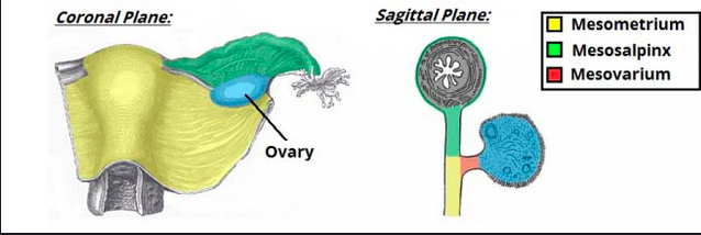
- Mesometrium – Surrounds the uterus and is the largest subsection of the broad ligament. It runs laterally to cover the external iliac vessels, forming a distinct fold over them. The mesometrium also encloses the proximal part of the round ligament of the uterus.
- Mesovarium – Part of the broad ligament associated with the ovaries. It projects from the posterior surface of the broad ligament and attaches to the hilum of the ovary, enclosing its neurovascular supply. It does not, however, cover the surface of the ovary itself.
- Mesosalpinx – Originates superiorly to the mesovarium, enclosing the fallopian tubes.
The three parts of the broad ligament.
Anatomical Relations
The broad ligament is related to many structures within the female pelvis. It is attached to the uterus, fallopian tubes and ovaries. These organs are supplied by the ovarian and uterine arteries, which are also contained within the broad ligament.
Three other ligaments of the female reproductive tract are located within the broad ligament:
- Ovarian ligament.
- Round ligament of uterus.
- Suspensory ligament of ovary (also known as the infundibulopelvic ligament).
(These ligaments shall be explored in more detail later in the article).
Ligaments Associated with the Ovary
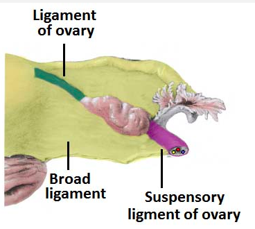
There are two main ligaments that attach to the ovary – the ovarian ligament and suspensory ligament of ovary.
Ovarian Ligament
The ovarian ligament is attached to the ovary inferiorly. It connects the ovary to the side of the uterus. Structurally, it is a fibrous band of tissue that lies within the broad ligament. It joins the uterus just below the origin of the fallopian tubes.
Proper ovarian ligament – Arvigo
Attaches the medial aspect of the ovary to the lateral aspect of the fundus
Suspensory Ligament of Ovary
The suspensory ligament of ovary extends outwards from the ovary to the lateral abdominal wall. It consists of a fold of peritoneum, thus some sources consider it to be part of the broad ligament. The function of this ligament is to contain the ovarian vessels and nerves (ovarian artery, ovarian vein, ovarian nerve plexus and lymphatic vessels).
Suspensory ligament of the ovary – Mercier
This is rounded cord like thickening of the broad ligament,located at its lower end, where it is attached to teh uterus., attaching to the ovaries and hold them in position.
Suspensory ligament of the ovary – Arvigo
These contain the arteries, veins, and lymph vessels and lymph vessels m and have the most tortorus path of all. They attach to the lateral wall of just under the fallopian tube, pass superolaterally (upward) to then attach at the iliac fossa, moving retro peritoneally (behind the peritoneal sac)and finally ending at L-3 with fascial connections.
Ligaments Associated with the Uterus
There are a number of ligamentous structures that attach to the uterus. They can be divided by where they attach to the uterus:
- Superior aspect – supported by the broad ligament and the round ligaments.
- Middle aspect – supported by the cardinal, pubocervical and uterosacral ligaments.
The inferior aspect of uterus is supported by the structures in the pelvic floor – the levator ani, perineal membrane and perineal body.
Round Ligament
The round ligament is a remnant of the embryonic gubernaculum. It originates at the uterine horns (the points at which the fallopian tubes enter the uterus), and attaches to the labia majora, passing through the inguinal canal. The round ligament can be a source of pain during pregnancy, due to the increased force placed on the ligament by the expanding uterus.
Round – Mercier
Starts at the top of the uterus starting at the top of the uterus and runs down the front of the abdomen. It then goes through the inguinal canal bikini line. The round ligament holds the uterus over the bladder tipped forwards oranterior. It also connects the uterus to the abdominal wall. This holds the uterus in place and gives pregnant woman some extra abdominal support during pregnancy.
ROUND – Arvigo.
Are situated between the layers of the broad ligments. They are 4-5 inch rounded fibromuscular cords, attaching from the lateral aspect of the fundus. passing anterior. They continue laterally through the internal abdominal ring, along and through the inguinal canal, and connect to the superifical perienal fascia at the labia where they extend like fingers.
Cardinal Ligaments
The cardinal ligaments are also known as the lateral, transverse cervical, or Mackenrodt’s ligaments. They are situated along the inferior border of the broad ligament and house the uterine artery and uterine veins. These ligaments arise from the side of the cervix and the lateral fornix of the vagina. They provide an extensive attachment on the lateral pelvic wall at the level of the ischial spines. Some fibres of the cardinal ligaments interdigitate with fibres from the uterosacral ligaments. When a hysterectomy is being performed due to a malignancy, the cardinal ligaments are often removed as they are common reservoir of cancerous cells.
CARDINAL – Mercier
The structure is situated at the root of the broad uterine ligament. It runs externally, and is continuous with the fibrous tissue that envelops the pelvic blood vessels.. It is primarily responsible for making a connection between the cervix and the lateral pelvic wall, which is located in the ischial spine and supports the uterus.
It also comprises of the uterine vein and the uterine artery.
During a radical hysterectomy is it essential to remove this ligament through surgical means.
Cardinal ligament – Arvigo
This is the principle ligament supporting the uterus and is the strongest in the body. it attaches in a circular manner round the cervix, moving literally, and connects to the obdurator fascia. it contains many of the blood vessels that come from the internal iliac artery.
Pubocervical Ligaments
The pubocervical ligaments are bilateral structures, which attach the cervix to the posterior surface of the pubic symphysis. They function to support the uterus within the pelvic cavity.
Pubo cervical ligament – Arvigo
Passes through the lateral inferior aspect of the uterus to surround the bladder and finally attach to the pubic symphysis.
Uterosacral Ligaments
THE move UTERINE RAKE SHORTENS THIS – allowing the prior lumbar back ache to no longer be telling you there is a problem. FIXES it.
The uterosacral ligaments are also bilateral fibrous bands, which attach the cervix to the sacrum. They are also known as the recto-uterine ligaments or sacrocervical ligaments. This supports the uterus and holds it in place.
UTEROSACRAL LIGAMENTS – Mercier
Is a paired ligament of the pelvis that runs between the uterus and the sacrum and the bone at the base for the vertebral column in the pelvis.
Also known as the retro-uterine ligament, it is one of the three paired peritoneal ligaments assisting in holding the uterus in place in the pelvic cavity. This ligament arises from the recto- uterine folds of the recto – uterine pouch, a space found just behind the cervix near the bottom of the uterus, and attaches to the front aspect of the sacrum.
As a dense band of connective tissue, the utero sacral ligament is largely made up of collagen fibres. It is essentially a combination of the recto-uterine folds bordering the lower uterus. The recto- uterine folds are made up of tissue from the peritoneum, (itself a membrane encapsculating the abdominal cavity, that extends downwards into the pelvis, ending where it meets the uterus along its anterior, superior and posterior borders.
UTEROSACRAL LIGAMENTS – Arvigo
Attach from the posterolateral aspect of the uterus, reaching around either side of the colon to attach to their posterior ends to the second and third sacral vertebral (anterior (Their attachments corresponds to the position of the uteroscaral junctions at the superficial aspect of the neck of the uterus.
And we now begin . . .